Mobile CT for Premium Imaging in the community
MARS brings diagnostics closer to patients with exceptional spatial resolution and low radiation dose.
As mentioned in


The Product
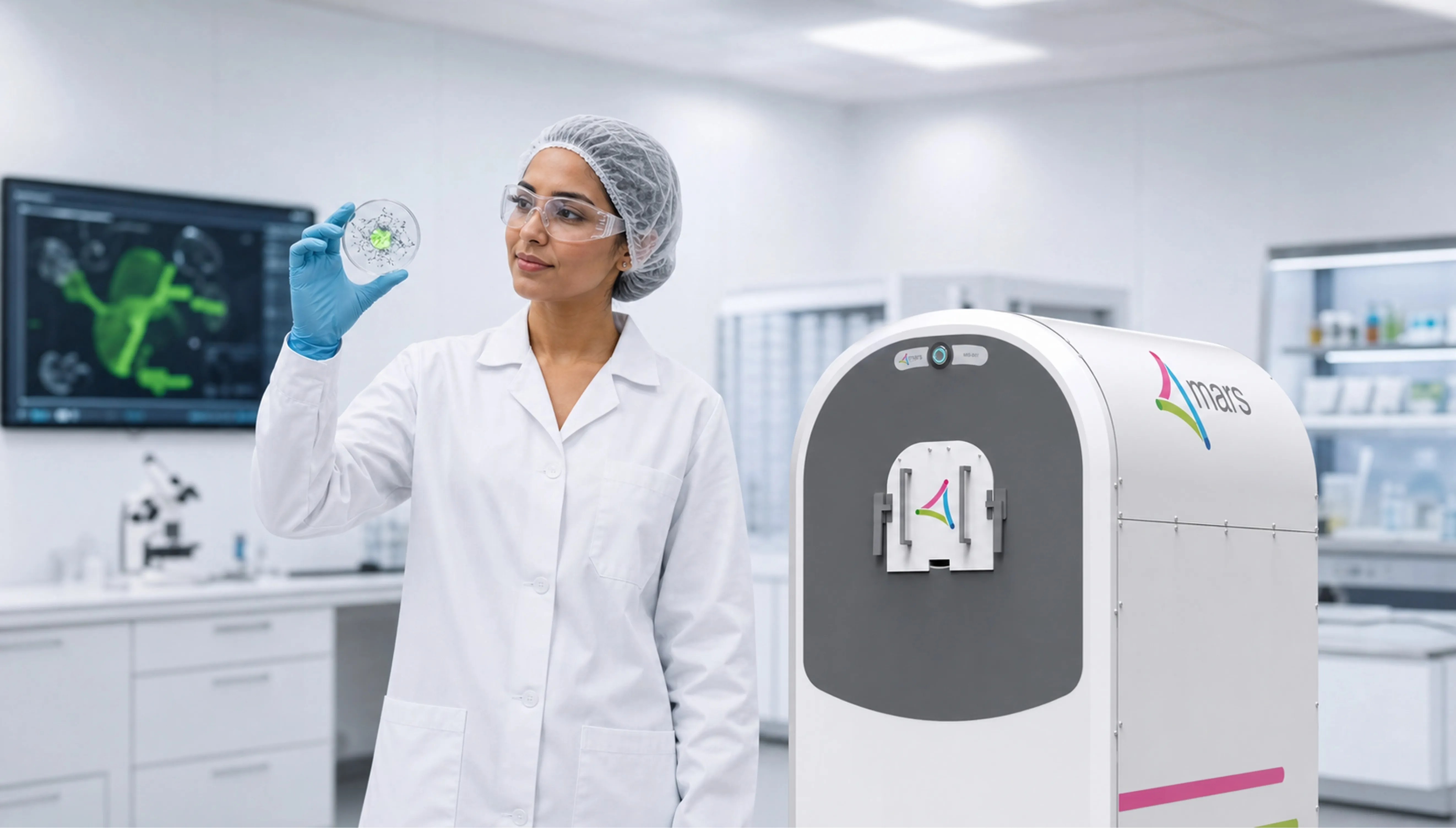
MARS Point-of-Care photon counting CT
Centralized CT facilities result in logistical delays, prolonged diagnosis, and higher costs. Advanced imaging systems are expensive to acquire and maintain, restricting access for smaller providers and raising per-scan expenses.
The MARS system delivers high-resolution extremity imaging with colour X-ray material detection, lower radiation exposure, and a compact footprint suitable for point-of-care use.
The Technology
Spectral Photon-counting CT
MARS photon counting detectors are capable of capturing and differentiating multiple x-ray energy levels, enabling identification and quantification of both intrinsic (e.g.,bone, soft tissue) and extrinsic (e.g.,contrast agents, nanoparticles) materials in a single scan.
Microlab
For pre-clinical spectral photon counting research
Specially designed for use in research laboratories, clearly see structure and get precise material identification and quantification all in one scan.
Case Studies
Clinical Research Collaboration
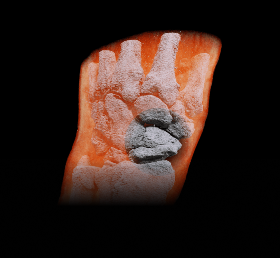
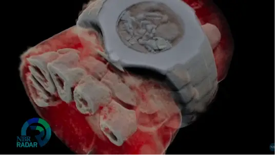
Scaphoid bone with sclerosis

Scaphoid fracture with sclerosis
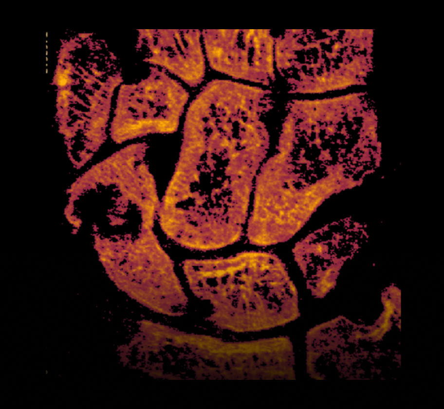
Non-union with displacement
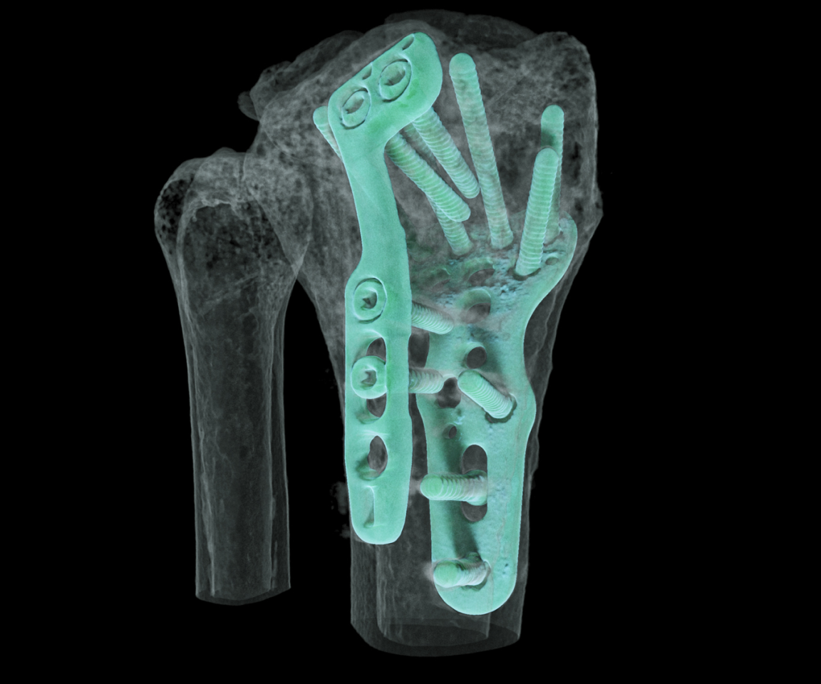
Scaphoid bone with sclerosis
Scaphoid fracture with sclerosis
Non-union with displacement
Our Team
A multidisciplinary team


Get in touch
Interested in clinical partnerships, technology collaborations, or learning more about the MARS system? Our team will respond shortly.
Christchurch Central City,
Christchurch, NZ 8011