Microlab Imaging System™
The world's first and only commercially available spectral photon-counting CT for preclinical research
We count individual photons and measure energy to see small anatomic structures and identify and quantify up to 6 materials with one scan, all in your regular lab environment.
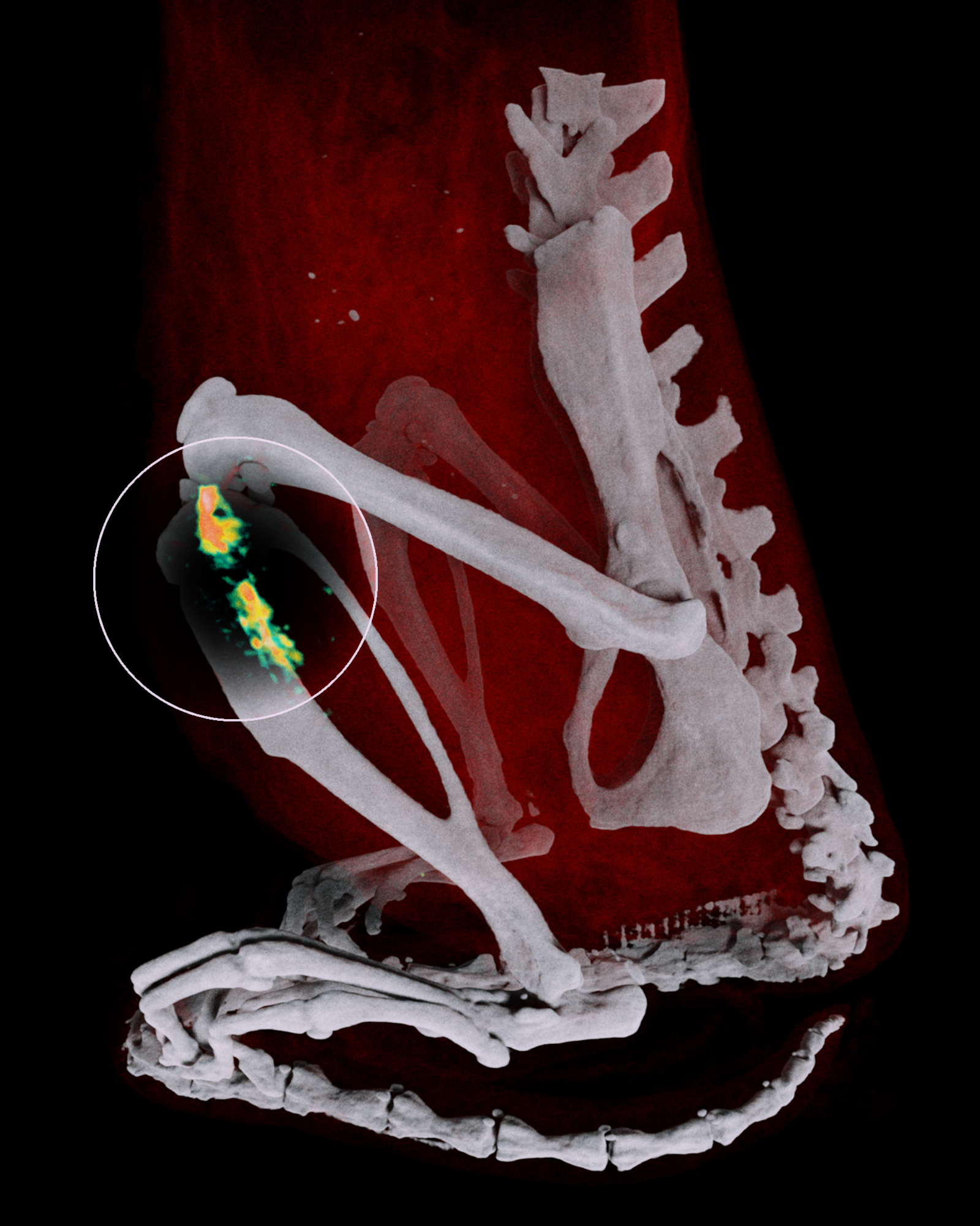
Research needs more than anatomy.
See what materials are present, at what concentration, simultaneously. Differentiate multiple contrast agents, distinguish drug compounds from tissue and observe nanoparticle distribution.
For researchers
Spectral imaging that brings your study to life
Ultra-high resolution (UHR) or 33 lp/cm with 0.9mm isotropic voxels. 2D and 3D volume renders with vivid colour overlays. Energy and material map previews. Precise region-of-interest measurement and per-material quantification.

For facilities managers
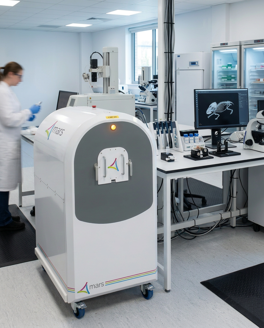
Built for the lab you already have
A fully shielded, mobile PCCT scanner that does not require a lead lined room or facilities alteration. Integrates with diagnostic imaging DICOM/PACS systems or data can be exported onto a standard Windows computer.
Technology & specifications
Microlab Imaging System™ uses Medipix3RX technology for advanced resolution of PCCT. Charge summing improves sensitivity and specificity. Metal artifacts reduced natively with features inherent to the system.
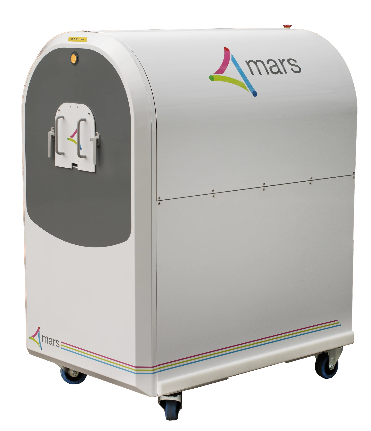
Technical specifications
- Detector
- CdTe PCD
- X-ray Tube
- 25 KHU
- Current
- 0.010 – 0.35 mA
- Bore
- 120 mm
- Footprint⁵
- 0.8 m² (8.6 ft²)
- Weight
- 405 kg (890 lb)
- Software
- MARS Scanner, MARS Vision

Contact us
Find out more
Discover how spectral imaging can transform your biomolecular research pathway.